Compact Bone Diagram : Seeley S 6 3 Bone Histology Pt 2 Compact Bone Diagram Quizlet / Compact bone, also called cortical bone, is the hard, stiff, smooth, thin, white bone tissue that surrounds all bones in the human body.
Compact Bone Diagram : Seeley S 6 3 Bone Histology Pt 2 Compact Bone Diagram Quizlet / Compact bone, also called cortical bone, is the hard, stiff, smooth, thin, white bone tissue that surrounds all bones in the human body.. Also called cortical bone, the compact variety usually features a haversian system, or cylindrical unit within the structure. Serves as protection of bone marrow. There are pores and spaces even in compact bone. Found in short bones, flat bones, irregular bones, and end of long bones osteons cylindrical structures that comprise compact bone, organized along lines of stress, constantly changing These bones are tough and hard with negligible gaps inside them.
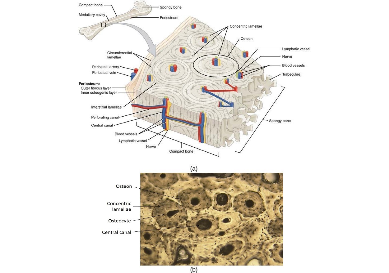
Haversian canals (sometimes canals of havers) are a series of microscopic tubes in the outermost region of bone called cortical bone. (b) in this micrograph of the osteon, you can clearly see the concentric lamellae and central canals. The compact bone gets its white, smooth structure owing to the connective tissues that cover around ¾ part of the bone from inside. Also called cortical bone, the compact variety usually features a haversian system, or cylindrical unit within the structure. The remainder is cancellous bone, which has a spongelike appearance with numerous large spaces and is found in the.
Compact bone diagram bone cross section diagram file624 diagram of compact bone new.
In compact bone, these cells are embedded within the solid calcium phosphate matrix of solid bone. It makes up the outer cortex of all bones and is in immediate contact with the periosteum. Compact bone is formed from a number of osteons, which are circular units of bone material and blood vessels. Some, mostly older, compact bone is remodelled to form these haversian systems (or osteons). Solution for diagram a limb bone, such as the tibia, showing the regionsof articular cartilage, spongy trabecular bone, compact bonetissue, and the marrow… Students view each slide and are asked to perform a task, like labeling or answering a question about the diagram shown. (b) in this micrograph of the osteon, you can clearly see the concentric lamellae and central canals. Diagram of a typical long bone showing both cortical (compact) and cancellous (spongy) bone. You need to get 100% to score the 10 points available. As compact bone grows, osteons begin to fuse together. Online quiz to learn compact bone diagram; Take a quick interactive quiz on the concepts in compact bone: Add to playlist 24 playlists.
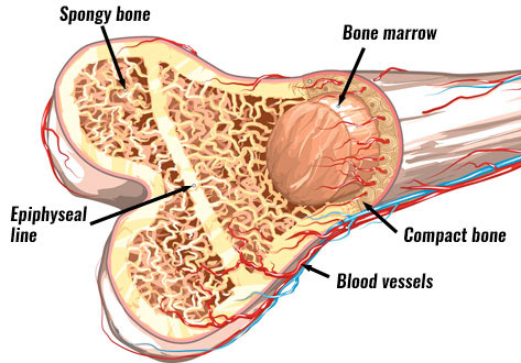
Compact and spongy tissues in a flat bone. They serve the purpose of protecting our bodies and also provide a structure and shape. (b) enlarged diagram of periosteum and compact bone in (a). These practice questions will help you master the. Diagram of a typical long bone showing both compact (cortical) and cancellous (spongy) bone.
You need to get 100% to score the 10 points available.
Serves as protection of bone marrow. Compact bone diagram osteon compact bone ap pinterest anatomy human anatomy and. You need to get 100% to score the 10 points available. Haversian canals (sometimes canals of havers) are a series of microscopic tubes in the outermost region of bone called cortical bone. Compact bone is formed from a number of osteons, which are circular units of bone material and blood vessels. Compact and spongy.the names imply that the two types differ in density, or how tightly the tissue is packed together. A diagram of the anatomy of a bone, showing the compact bone. They allow blood vessels and nerves to travel through them to supply the osteocytes. Compact bone diagram bone cross section diagram file624 diagram of compact bone new. With students learning from home, i needed to get creative with how students could learn how bone is organized and how it grows and remodels. Compact bone, also called cortical bone, dense bone in which the bony matrix is solidly filled with organic ground substance and inorganic salts, leaving only tiny spaces (lacunae) that contain the osteocytes, or bone cells.compact bone makes up 80 percent of the human skeleton; Diagram of a typical long bone showing both cortical (compact) and cancellous (spongy) bone. As compact bone grows, osteons begin to fuse together.
Add to playlist 24 playlists. As seen in the image below, compact bone forms the cortex, or hard outer shell of most bones in the body. In development there are 2 separate signaling pathways for pattern formation and the formation of bone itself. The remainder is cancellous bone, which has a spongelike appearance with numerous large spaces and is found in the. Compact bone diagram bone cross section diagram file624 diagram of compact bone new.

Solution for diagram a limb bone, such as the tibia, showing the regionsof articular cartilage, spongy trabecular bone, compact bonetissue, and the marrow…
Take a quick interactive quiz on the concepts in compact bone: As seen in the image below, compact bone forms the cortex, or hard outer shell of most bones in the body. Compact bone, as opposed to spongy bone, is made of cylindrical units, called osteons, that are tightly formed together. The shafts found in long bones are also compact bones. The key difference between compact bone and spongy bone is that the compact bone is a tough and heavy bone that forms the diaphysis of long bones while the spongy bone is a soft and light bone that forms the epiphysis of long bones. The compact bone gets its white, smooth structure owing to the connective tissues that cover around ¾ part of the bone from inside. Found in short bones, flat bones, irregular bones, and end of long bones osteons cylindrical structures that comprise compact bone, organized along lines of stress, constantly changing Online quiz to learn structure of compact bone; Compact bone diagram bone cross section diagram file624 diagram of compact bone new. Human body thoracic diagram 7 photos of the human body thoracic diagram abdomen human body, chest human body, human thorax diagram, sternum human body, thoracic cavity diagram, thoracic diaphragm, thoracic nerve diagram, human anatomy, abdomen human body, chest human body, human thorax diagram, sternum human body. A diagram of the anatomy of a bone, showing the compact bone. Terms in this set (8) spongy bone (contains red marrow) compact bone (has osteons) osteon. It is also called osseous tissue or cortical bone and it provides structure and support for an organism as part of its skeleton, in addition to being a location for the storage of minerals like calcium.about 80% of the weight of the human skeleton comes from.


Komentar
Posting Komentar